Anatomical Heart’s Hidden Secrets
Anatomical Heart’s Hidden Secrets goes through the unseen details of the human heart, which is usually seen as a simple pump for our body. This guide tells surprising truths about its design, job and mysteries hidden within it. From the complexities in the electrical system to unexpected features of blood flow, this tour reveals a new viewpoint on an organ we often overlook.
Anatomical Heart’s Location and Size
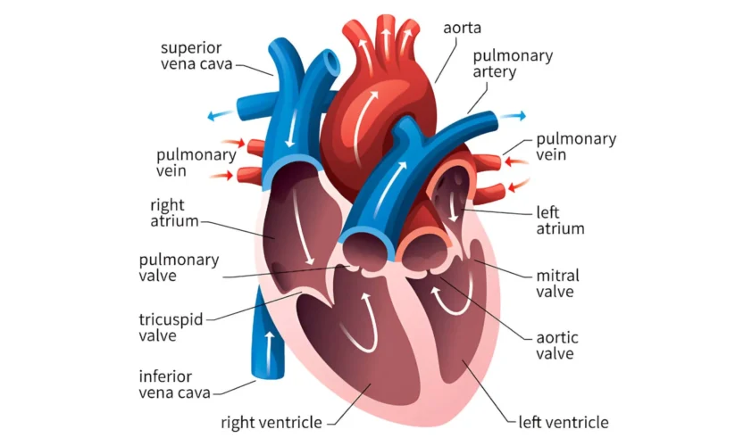
The anatomical heart is found in the thoracic cavity, which means it’s in the chest area between lungs and a little to the left side. Its size can be likened to that of someone’s hand when they make a fist.
The Heart’s Structure
We can think of the anatomical heart as a busy house with four rooms. The two smaller rooms on top, called atria, get blood from different parts of our body. Then we have the bigger lower chambers which are known as ventricles; these act like strong pumps pushing out oxygenated blood into arteries for delivering it throughout our system again while also receiving deoxygenated blood back from veins to complete this loop process once more time.
The anatomical heart’s doors or valves are present between these rooms: they open and shut very precisely to guide flow direction in an orderly manner. The heart’s walls, like strong building structures, maintain the function of this amazing organ.
The Anatomical Heart’s Electrical System
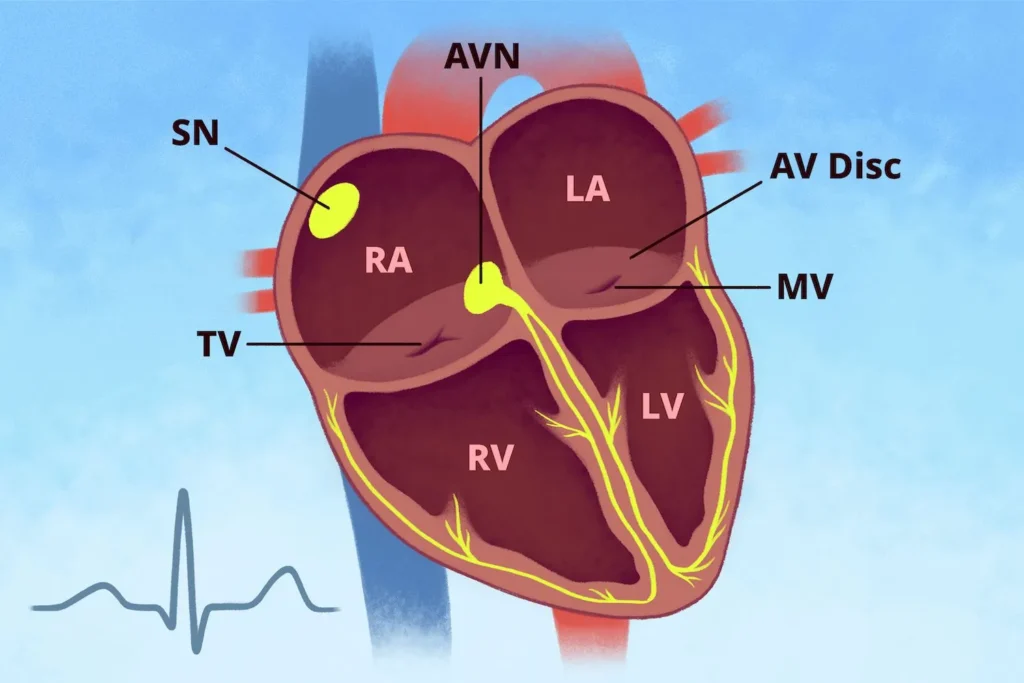
The electrical system of the heart is similar to a cheering crowd present in a stadium. It begins with a SA node, which can be compared to an energetic cheerleader who starts the applause. This wave of excitement or electrical impulse spreads through atria that are like upper stands.

The AV node, similar to a gatekeeper, somewhat slows down the cheer so that all can be ready. Then the Bundle of His and its branches, like cheerleaders spreading enthusiasm through a wave, carry this excitement to ventricles (lower stands).
At last Purkinje fibers make sure every seat feels thrilled just like most energetic fans in the front row.
The Heart’s Blood Supply
Think about the anatomical heart like an athlete that is very active. Just as with any muscle, it needs a continuous flow of nutrients and oxygen for working at its best. These important elements are brought to the heart muscle by coronary arteries, similar to rivers giving life.
Like tributaries branching out, they make sure that each part of the heart gets what it needs. On the other hand, coronary veins function like drainage channels to remove waste products.
The Heart’s Work
The anatomical heart functions in a manner similar to a rhythmic pump. It goes through periods of tightening (systole) and loosening (diastole), much like the squeezing and releasing of a toothpaste tube. This action is comparable to how ventricles contract then relax, resulting in blood being pushed out from them.
Next, letting go of the force is similar to diastole. It permits the chambers to be filled again. This joint movement pushes blood through the body’s extensive system of vessels. To truly comprehend this complex process, viewing animations or videos showcasing the cardiac cycle proves extremely useful.
The Heart’s Valves
Think of the valves in the heart like smart doorkeepers. The tricuspid valve, which is a three-door guard, manages what goes in and out between the right atrium and ventricle. Pulmonary valve acts similar to a one-way exit letting blood travel towards the lungs.
The mitral valve, having two doors, is responsible for controlling blood flow between the left atrium and ventricle. At last, we came to the aortic valve on our grand exit. It pushes oxygen-filled blood out into our body.
These are like vigilant soldiers making sure that blood moves only in one direction, not allowing any undesirable reversals.
The Heart’s Layers
The wall of the anatomical heart has three layers, and each layer does a special job. The epicardium is like a bodyguard that coats the outside of the heart; it protects this important organ from harm as if it’s in an embrace.
Below this protective layer is muscle power called myocardium, which makes sure the heart contracts in rhythm. It’s like a hardworking athlete, pushing blood around the body.
The endocardium, which is a smooth inner lining, makes sure that blood moves without friction. It can be compared to how a pipe looks inside when it gets polished and well maintained just as this allows for easy travel of fluid in the form of life giving liquid, so too does endocardium facilitate smooth journey for blood through the human body.
The Heart’s Neighbors
The pericardium, which is a double-layered sack filled with fluid, surrounds and safeguards the heart. It works as a shock absorber for the heart’s movements while it beats. The pericardium also aids in keeping the heart steady within its location in your chest cavity.
The anatomical heart’s position within the chest, specifically in the middle area between lungs, is significant and intricate. It is divided from the lungs by a region named mediastinum which holds not only heart but also trachea, esophagus and main blood vessels.
The pericardium, a double-layered sac, is responsible for safeguarding the heart and keeping it in place inside the chest cavity. The outer part of this sac that surrounds and protects our hearts is known as the parietal pericardium. It links with the wall of our chests.
Anatomical Heart’s Blood Vessels
In your physical body, there are three major kinds of blood vessels: arteries, veins and capillaries. Arteries transport blood from the heart to other tissues in your body. Veins carry it back to the heart again.
Capillaries are very small vessels which link up with both arteries and veins in an intricate network throughout your system. They function as the location where oxygen, nutrients, and waste products are traded between blood and cells in the body.
The movement of blood from the heart to lungs and back again is called “pulmonary circulation.”
Blood going around the body, also known as “systemic circulation,” moves from heart to all parts of our body before it goes back into heart. In the pulmonary arteries, blood takes oxygen from lungs and releases carbon dioxide.
Blood that is filled with oxygen now goes back to the heart through pulmonary veins. The heart pushes blood into the body’s tissues using systemic arteries, where it gives away oxygen and nutrients. Blood without oxygen comes back to the heart through systemic veins.
Keeping Your Heart Sweet
An anatomical heart that is in good health will also be happy! You can keep your heart healthy by eating a diet which has balance, low amounts of saturated and trans fats, cholesterol and sodium. Make an effort to do at least 30 minutes of exercise with medium intensity on most days every week.
Have enough sleep and deal with stress using methods like yoga or meditation. Visit your doctor for regular checkups and screenings, even when you feel healthy. Taking care of your heart can lower chances of heart disease and let you live more years in good health.
The anatomical heart is undoubtedly the engine of life. Its complicated operations such as rhythmic contractions or quiet electrical signals depict the intricacy and excellence found within our bodies.


